A 63-year-old woman who had presented to the ophthalmology clinic for evaluation of cataracts was found to have white-yellow rings in both eyes. Over the past few years, she had noticed mild worsening of her vision. She reported no history of keratitis or ocular trauma. A lipid panel had been normal at a health maintenance visit 6 weeks before presentation. On ophthalmologic examination, peripheral opacities were observed in the lens of each eye, a finding consistent with age-related cataracts. A funduscopic examination was normal. Visual acuity was 20/30 in each eye. On slit-lamp examination, two concentric white-yellow rings were seen in each cornea (Panel A shows the right eye, and Panel B the left eye). No corneal thinning or inflammation was apparent. A diagnosis of double corneal arcus was made. Corneal arcus (also known as arcus senilis) is an annular opacity that results from lipid deposition. It typically manifests as a single, peripheral ring but may rarely manifest as a double ring, as seen in this patient. Corneal arcus is a consequence of normal aging in older patients and may be associated with hypercholesterolemia in younger patients. The patient was counseled that the double corneal arcus was a benign finding and that her decreased visual acuity was related to the cataracts.
A healthy 72-year-old man who had emigrated from Zimbabwe 20 years previously presented to the dermatology clinic with a 17-year history of swelling of the penis, scrotum, and left leg. During a recent hospitalization, nonpitting edema of the left leg had been noted, and a urinary catheter had been placed temporarily (Panel A). At the current presentation, there was nonpitting edema of the penis and scrotum (Panel B), as well as ongoing swelling of the left leg. Laboratory testing showed an eosinophil count of 500 per cubic millimeter (normal range, 0 to 300). Magnetic resonance imaging of the pelvis showed swelling of the scrotal tissues on both sides; no hydrocele was present. An enzyme-linked immunosorbent assay and an indirect fluorescent antibody test for Wuchereria bancrofti were positive. A blood smear to identify microfilariae was not obtained. A diagnosis of chronic lymphatic filariasis — a mosquito-borne parasitic infection in which nematodes invade the lymphatic system — was made. The patient was treated initially with a course of doxycycline and single-dose albendazole. Single-dose diethylcarbamazine was given later, after tests for concomitant onchocerciasis and loiasis returned negative. Diethylcarbamazine administration is contraindicated in patients with lymphatic filariasis and concomitant onchocerciasis or loiasis owing to the risk of severe adverse reactions from the rapid killing of microfilariae. At follow-up 2 months after the completion of treatment, the patient’s symptoms had resolved.
Summary: Umbilical cord milking (UCM)—a method of squeezing the umbilical cord towards the infant before clamping—poses no long-term neurodevelopmental risks for newborns. The study involved assessing 971 children at ten U.S. medical centers, evaluating key developmental areas such as communication and motor skills.
Results indicate that children who underwent UCM at birth did not exhibit increased neurological challenges compared to those who received early cord clamping. This finding supports UCM as a viable, no-cost strategy for enhancing the health of newborns, especially in resource-limited settings.
Key Facts:
Safety of UCM Confirmed: Long-term follow-up shows that UCM does not increase the risk of neurodevelopmental challenges in children.
Broad Evaluation: The study assessed multiple developmental aspects, including motor and social skills, across a large cohort of children.
Implications for Low-resource Settings: UCM offers a cost-effective method to improve newborn health, particularly valuable in areas with limited medical resources.
Source: Pediatric Academic Societies
An alternative method of transferring blood cells to weakened newborns through their umbilical cord does not carry long-term neurodevelopmental risks compared to standard practice, a recent study found.
The research will be presented at the Pediatric Academic Societies (PAS) 2024 Meeting, held May 3-6 in Toronto.
Study authors found that children who received UCM at birth were no more likely to have neurological challenges two years after the procedure compared to those who received early cord clamping.
Umbilical cord blood contains oxygen and beneficial nutrients for newborns, experts say. Doctors may delay clamping a newborn’s umbilical cord to pass nutrients through their cord if they have poor breathing or a low heart rate immediately after birth.
A study found that umbilical cord milking (UCM), an alternative method of transferring cord blood where a doctor squeezes the umbilical cord toward the infant before clamping, does not cause long-term harm.
Researchers assessed 971 children’s communication, motor skills, problem-solving, and social skills across 10 U.S. medical centers. Study authors found that children who received UCM at birth were no more likely to have neurological challenges two years after the procedure compared to those who received early cord clamping.
“The short- and long-term benefits point to UCM as a safe alternative for ensuring weakened newborns can live a full, healthy life,” said Anup Katheria, MD, physician at Sharp Mary Birch Hospital for Women & Newborns and presenting author.
“UCM is a no-cost option for obstetricians to preserve the health and wellbeing of children.”
Study authors say raising awareness of UCM as a safe option for supporting weakened newborns is important to promoting equity among resource-limited settings.
The U.S. Preventive Services Task Force recommendation comes as more women are being diagnosed with breast cancer at a younger age.
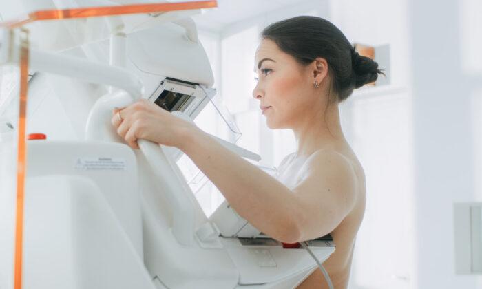
A national advisory panel has reduced the recommended age for breast cancer screening, suggesting women get their first mammogram at age 40 instead of 50 and continue every other year until age 74.
The U.S. Preventive Services Task Force recommendation comes as more women are being diagnosed with breast cancer at a younger age. It is a reversal of the panel’s previous recommendation suggesting women make an individual choice about getting a mammogram between ages 40 and 49. The recommendation applies to women at average risk of breast cancer, as well as those with a family history of breast cancer and those with dense breasts.
“More women in their 40s have been getting breast cancer, with rates increasing about 2 percent each year, so this recommendation will make a big difference for people across the country,” Task Force Chair Dr. Wanda Nicholson said in a press release. “By starting to screen all women at age 40, we can save nearly 20 percent more lives from breast cancer overall.”
The task force published the recommendations in 2023 as a draft open for public comment. The draft was finalized on Tuesday, April 30, and published in the Journal of the American Medical Association.
Mortality Rates Declining, but Breast Cancer Rates Increasing
According to the American Cancer Society, breast cancer is the second leading cause of cancer death for women in the United States, despite a steady decline in breast cancer mortality over the past 20 years. Most cases occur in women between the ages of 55 and 74, with the highest death rates occurring in women with a median age of 70.
Along with younger women, the new guidelines aim to help black women, who are 40 percent more likely to die of breast cancer than white women, according to Dr. Nicholson, who added that the new guidelines are just another step toward improving existing inequalities in the American health care system.
“We need to know how best to address the health disparities related to breast cancer so all women can live longer and healthier lives,” added Dr. John Wong, vice chair of the task force. “Clinicians must help reduce any barriers to patients getting the recommended screening, timely, equitable, and appropriate follow-up, and effective treatment of breast cancer.”
More Room for False Positives?
Adding another decade of testing increases the risk of experiencing at least one false positive during a mammogram, according to a 2022 University of California (UC)–Davis Health study published in JAMA Network Open. The study found that half of all women will experience at least one false positive over a decade of annual breast cancer screening. However, the risk of a false positive was considerably lower if screening occurred every other year (as recommended by the U.S. Preventive Services Task Force) as opposed to annually over a decade.
False positives are also more likely for women with denser breast tissue. The task force noted that more research is needed to show how screening with breast ultrasound or MRI might better help women with dense breasts.
“Findings from our study highlight the importance of patient-provider discussions around personalized health. It is important to consider a patient’s preferences and risk factors when deciding on screening interval and modality,” Michael Bissell, co-first author of the UC Davis Health study, said in a 2022 news release.
False positives are common. Although only 12 percent of 2D screening mammograms require more testing, less than 1 percent result in a cancer diagnosis, according to the UC Davis press release. Not only can they be expensive and timely, but they can also cause the patient to undergo unnecessary stress.
“Despite the important benefit of screening mammography in reducing breast cancer mortality, it can lead to extra imaging and biopsy procedures, financial and opportunity costs, and patient anxiety,” Diana Miglioretti, professor and division chief of biostatistics at UC Davis’ Department of Public Health Sciences, said in the 2022 press release.
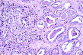
A novel treatment for leukemias and lymphomas that arise from immune system T cells, developed by investigators at the Johns Hopkins Kimmel Cancer Center and its Ludwig Center and Lustgarten Laboratory, was found to be effective at killing these cancers in mice bearing human T-cell tumors.
The therapy, an antibody-drug conjugate (ADC), combines an antibody that targets a protein called TRBC1 expressed on the surface of T-cell cancers with an anti-cancer drug, called SG3249. The ADC works by using the antibody to seek out the cancer cells that express TRBC1. Then, those cancer cells ingest the ADC, where SG3249 is released and kills the cancer cells. A description of the work was published in Nature.
Each year, about 100,000 patients worldwide are affected by T-cell leukemias and lymphomas. Adults with relapsed T-cell cancers have limited therapeutic options and five-year survival rates of 7–38%.
“Developing treatments for T-cell leukemias and lymphomas is much more difficult than for leukemias and lymphomas arising from immune system B cells,” explains senior study author Suman Paul, MBBS, PhD, an assistant professor of oncology at the Johns Hopkins University School of Medicine. Effective therapies for B-cell cancers wipe out both cancerous and noncancerous B cells, but patients still do well even without the immune system B cells that help fight infections. However, if similar approaches are used and a treatment wipes out both normal and cancerous T cells, it would leave patients without a functioning immune system and at high risk of dying from infections.
“Not much drug development has happened in this space of T-cell leukemias and lymphomas,” Paul says. “We need new therapies for these cancers, but whatever therapies we develop in the space have to be cancer-specific. We have to preserve some of the normal T cells and wipe out cancerous T cells at the same time.”
T-cell cancers express either TRBC1 or TRBC2, while normal T cells express a mix of TRBC1 and TRBC2. Therefore, selective targeting of TRBC1 can potentially eradicate the normal and cancerous T cells expressing TRBC1 while preserving normal T cells expressing TRBC2. A recent clinical trial conducted elsewhere attempted to target TRBC1 cancers using chimeric antigen receptor (CAR) T-cell therapy. These CAR T cells are genetically engineered T cells that bind to and kill TRBC1 cells. CAR T-cell therapies are FDA-approved treatment options used in several B-cell cancers. However, after administering the TRBC1-targeting CAR T cell therapy in human patients, trial investigators reported that the CAR T cells were not persisting inside the patients. Such persistence is required for effective cancer cell-killing. Interested to understand why, Paul and colleagues found that the CAR T cells targeting TRBC1 could be killed by normal T cells, limiting their persistence.
This lack of CAR T-cell persistence led the team to try TRBC1 targeting with the use of antibody-drug conjugates. Paul and colleagues tried two different formulations of ADCs in mouse models of T-cell cancers. After a single injection of one formulation of the treatment, the cancers initially regressed but then recurred. After a single treatment with the anti-TRBC1-SG3249 ADC combination, investigators observed signs of cancer elimination within seven days and the cancers were eventually undetectable, with no recurrences. “The tumors didn’t come back, and we followed the mice for more than 200 days,” Paul explains.
The treatment was able to eliminate the cancer while preserving half of the remaining normal T cells. “The residual normal T cells should be sufficient to maintain some immune system protection against infectious diseases,” Paul says.
“Witnessing the successful elimination of T-cell cancers while sparing normal T cells in preclinical models was truly gratifying,” adds Jiaxin Ge, a co-author of the study and third-year Ph.D. student in the Ludwig Center. “We believe this approach has the potential to address a critical unmet need in oncology, and we’re committed to advancing it through further research.”
Tushar Nichakawade, first author on the study and a fourth-year PhD student at the Ludwig Center, says, “There are so many lessons to learn from the clinic and it has been exciting to be a part of the iterative process of drug discovery. Every therapy has its pros and cons, but the preclinical efficacy of our ADC gives me hope that it can make a difference for patients suffering from these terrible cancers.”
Biomedical imaging modalities such as magnetic resonance imaging (MRI) have revolutionized the ability to detect and track the progress of many cancer types. However, the difficulty of obtaining detailed images of cancer cells buried deep within normal tissues has slowed the usefulness of imaging technology for improved, personalized cancer care.
An approach biomedical engineers are pursuing to overcome this limitation involves building nanoprobes that are designed to travel throughout the body, accumulate in cancer cells, and send a signal that illuminates the tumor.
NIBIB-supported researchers at Johns Hopkins University have developed a smart nanoprobe designed to infiltrate prostate tumors and send back a signal using an optical imaging technique known as Raman spectroscopy. The new probe, reported in Advanced Science, has the potential to determine tumor aggressiveness and could also enable sequential monitoring of tumors during therapy to quickly determine if a treatment strategy is working.
“We are excited about the potential for improving diagnosis and treatment of many common cancers,” explained the project leaders Jeff Bulte, PhD, and Ishan Barman, PhD. “We are combining self-assembling nanoprobes with Raman spectroscopy to achieve precise, single-cell resolution images required for eventual practical use in the clinic.”
The engineering team constructed a nanoprobe that is sensitive to its local microenvironment: it is activated only after it encounters legumain, a tumor-associated enzyme that is produced by aggressive prostate cancer cells. Once it encounters the nanoprobe, legumain splits it into pieces that can self-assemble to create an optically active nanoparticle. These nanoparticles emit specific wavelengths of light that can be detected with Raman spectroscopy to visualize the tumor.
Postdoctoral fellows Swati Tanwar, PhD, and Behnaz Ghaemi, PhD, spearheaded the design and synthesis of the nanoprobe, called nanoSABER (for Self-Assembling Bioorthogonal Enzyme Recognition)—an apt name that reflects the surgical precision of the smart molecule.
“We have chosen Raman reporters that are specifically active in the ‘cell silent’ region of the near-infrared spectrum to avoid interference with the signal from normal tissue,” explained Barman. “This selective activity is crucial for our imaging technique, as it allows for precise detection by Raman spectroscopy without reacting with or being obscured by the surrounding biological material.”
The researchers used two prostate cancer cell lines exhibiting varying levels of legumain expression—one high and one low—alongside a non-cancerous prostate epithelial cell line, which produces a negligible amount of the enzyme.
NanoSABER was tested in laboratory cell cultures and in an experimental mouse model. In both settings, the prostate cells expressing legumain activated the nanoprobe and emitted a signal with an intensity that corresponded to the amount of legumain produced by the cancer cells. The non-legumain cell type did not activate the nanoprobe, demonstrating that the nanoSABER system performed as designed, emitting a signal that correctly indicated the presence and amount of the cancer-associated enzyme.
“The work by this group is a significant step towards better care for men afflicted with prostate cancer,” explained Tatjana Atanasijevic, PhD, a program director in the Division of Applied Science & Technology at NIBIB. “It is an excellent example of the type of innovative technologies NIBIB supports that have the potential to dramatically impact health care.”
The team believes they have engineered a molecular system with the potential to not only identify tumors using optical imaging but to also rapidly assess tumor aggressiveness—potentially without the need for painful biopsies that are the current standard of care. In addition, as profiles of enzyme secretion by different types of cancers are discovered, additional nanoSABER probes can be synthesized that will allow a level of precise diagnosis of tumor types and characteristics that is not currently possible, including sequential imaging of tumors to determine whether therapies are working in real time.
SYNC-T, an investigational therapy that combines a device-induced vaccination at the tumor site with intratumoral infusion of a multitarget biologic drug, led to numerous clinical responses in patients with metastatic castrate-resistant prostate cancer (mCRPC). The results were reported at the American Association for Cancer Research (AACR) Annual Meeting 2024.
Patients with mCRPC—prostate cancer that does not respond to hormone therapies—have few treatment options and a high mortality rate, according to Charles Link, MD, an adjunct professor at Lankenau Institute for Medical Research, part of Main Line Health, and a cofounder and executive chairman of Syncromune. Prostate cancer has an immunologically “cold” tumor microenvironment. This has presented challenges for existing immunotherapies, which have exhibited low response rates and high toxicity in patients with prostate cancer.
Link and colleagues developed a novel treatment approach to stimulate a systemic antitumor immune response for mCRPC. Their therapy, called SYNC-T, first uses a probe that is inserted directly into the primary or metastatic tumor to freeze a portion of the tumor, which causes the tumor cells to fracture (oncolysis) and release immune-stimulating neoantigens. In essence, this method generates a personalized in situ neoantigen cancer vaccine that serves to activate the immune system, Link explained.
Link added that the imaging and procedural techniques for inserting a probe into the prostate are similar to the methods routinely used by urologists to conduct prostate biopsies. Immediately following the oncolysis step, an investigational multitarget biologic drug called SV-102—which is a fixed-dose drug comprised of active pharmaceutical ingredients: an anti-PD-1 antibody, an anti-CTLA4 antibody, a CD40 agonist, and a TLR9 agonist—is infused into the area of lysis in the tumor.
“SV-102 simultaneously blocks two distinct mechanisms of immune suppression and activates two distinct mechanisms of immune enhancement, allowing the vaccine-induced T cells to activate and mount a systemic antitumor immune response,” said Link.
The safety and efficacy of SYNC-T were evaluated in a phase I clinical trial, which enrolled 15 patients (12 patients with mCRPC and three patients with metastatic prostate cancer who opted out of hormone therapy), 13 of which have been evaluated for response. Sixty percent of patients identified as white, 33% as Hispanic, and 7% as Black. The median age was 61 years.
Of the 13 evaluable patients, 11 experienced an objective response, with five complete responses and six partial responses). The other two evaluable patients had stable disease at the time of data analysis. Six patients experienced mild to moderate treatment-related adverse events, including fever, rigors, fatigue, diaphoresis, hematuria, urinary tract infection, acute urinary retention, and hepatic enzyme elevation.
“The toxicity of SYNC-T appears to be much lower than what has been observed previously with intravenous immunotherapy for prostate cancer,” Link noted.
“Our results indicate that SYNC-T is associated with a high response rate without generating severe toxicity in this initial group of patients, which opens up opportunities to expand the role of immunotherapy in mCRPC,” he summarized. “Further, this approach uses standard procedures already employed by urologists and radiologists, which means the treatment could potentially be rapidly adopted by treating physicians.”
Limitations of the study include the small sample size, short follow-up time, and the single-arm design.
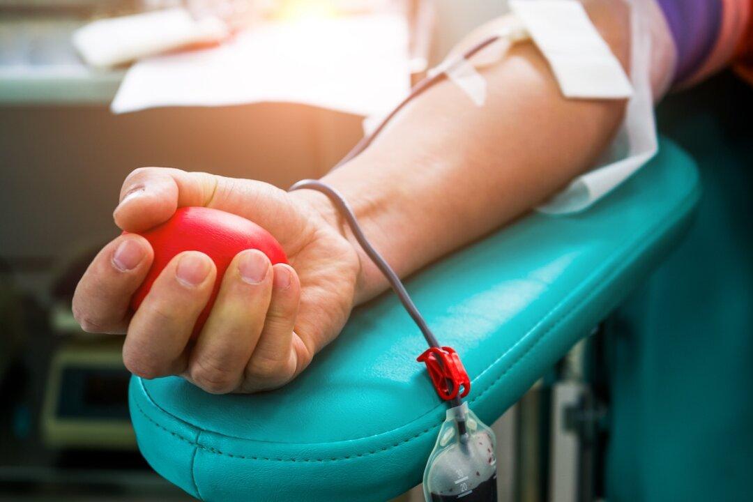
An enzyme discovered in the human gut can remove antigens from A, B, and AB blood to make it into universal donor blood, a new study showed.
Scientists at the Technical University of Denmark (DTU) and Lund University have gotten one step closer to cracking the code to making universal donor blood after discovering enzymes that can remove specific sugars comprising the A and B antigens in A, B, and AB blood types when the enzymes are mixed with red blood cells.
The study, published in Nature Microbiology on April 29, also offers insight into each blood type’s unique structures.
“Universal blood will create a more efficient utilization of donor blood, and also avoid giving [ABO]-mismatched transfusions by mistake, which can otherwise lead to potentially fatal consequences in the recipient,” professor Martin L. Ollson, leader of the study at Lund University, said in a press release. “When we can create [ABO]-universal donor blood, we will simplify the logistics of transporting and administering safe blood products, while at the same time minimizing blood waste.”
Blood donation is a constant need in the United States. The American Red Cross reports that a person needs blood in the United States every two seconds. Donated blood is required for surgeries, cancer treatment, chronic illnesses, and traumatic injuries. The organization reported a blood donation shortage at the start of 2024, noting that blood donations through the Red Cross have fallen by about 40 percent over the past two decades. Some reasons for this include the COVID-19 pandemic driving remote work, donor eligibility changes, and revisions to blood transfusion protocol in hospitals.
How Enzymes Can Help Create Universal Donor Blood
The idea of enzymes being used to make blood has been around for over 40 years. The theory is that the enzymes work as they would in the stomach, eating away the sugar structures in the blood that make it type A, B, or AB. The sugar structures of each of these three blood types are called antigens; if these antigens are mixed with incompatible blood, it can trigger a dangerous immune response. Type O blood lacks any of these antigens, so it is possible to transfer this type to any person, regardless of their blood type.
According to the American Red Cross, only 7 percent of people in the United States have type O negative blood, which is why it is always in great demand and short supply. Consequently, developing a way to remove the A and B antigens from A, B, and AB blood types is highly desired to meet the demand.
Although researchers have been working with antigen-removing enzymes for four decades, and more efficient enzymes have been discovered, problems eliminating all immune reactions remain. Hence, the enzymes have yet to be used in clinical practice.
The research team at DTU and Lund University, however, discovered a new cocktail of enzymes extracted from the human gut bacterium Akkermansia muciniphila. This bacterium breaks down the mucus covering the gut’s surface, structurally similar to the complex sugars found at the surface of blood cells.
They tested 24 enzymes on hundreds of blood samples.
“What is special about the mucosa is that bacteria, which are able to live on this material, often have tailor-made enzymes to break down mucosal sugar structures, which include blood group [ABO] antigens. This hypothesis turned out to be correct,” professor Maher Abou Hachem, study lead at DTU and one of the senior scientists, said in a press release.
The research team admitted that, while the findings are promising, more work remains. They noted that they are close to perfecting universal blood from type B donors, but issues remain with type A blood, which is more complex.
“Our focus is now to investigate in detail if there are additional obstacles and how we can improve our enzymes to reach the ultimate goal of universal blood production,” Mr. Hachem said.
The study found that lipid nanoparticles cause inflammation by damaging cellular components called endosomes, but the degree of inflammation varies.
A recent preprint from the University of Pennsylvania (UPenn) showed that lipid nanoparticles, which transport COVID-19 mRNA in COVID-19 vaccines, cause inflammation within cells. However, this inflammation can be reduced if the proper lipid nanoparticle is used.
The researchers tested several different lipid nanoparticle formulations in animals, including ALC-0315 and SM-102, used in Pfizer’s and Moderna’s COVID-19 vaccines, respectively.
They found that all the tested lipid nanoparticles cause inflammation by damaging cellular components called endosomes. However, the degree of inflammation varies depending on the degree of damage.
Additionally, the researchers learned that a lipid nanoparticle called 4A3-SC8 induced a lower level of inflammation by causing less damage. Inflammation could also be reduced with the drug thiodigalactoside, which prevents the cell from detecting severe damage.
Since the rollout of the COVID-19 vaccines, lipid nanoparticles have become a popular technology in nanomedicine.
“Right now, the whole nanomedicine field is focused on how to amplify LNP (lipid nanoparticle) formulation for all other diseases,” Wang Yufei, a postdoctoral researcher at UPenn and one of the study’s lead authors, told The Epoch Times.
The researchers wrote in the preprint, “Our group and others have recently shown that LNPs can induce severe inflammation and worsen markers of pre-existing inflammation by up to [greater than] 10-fold.”
Most people who took the COVID-19 vaccines did not experience severe adverse reactions, but some papers have alluded to hyperinflammatory syndromes that occur post-vaccination.
“One question is, how can we use LNPs in a safer and more efficient way? So that is one of the main purposes that we try to optimize the LNP formulation and try to understand the mechanics of LNP interaction with the whole body,” Ms. Wang said.
Lipid Nanoparticles Punch Holes in Cell Structures
The authors also explored how lipid nanoparticles cause inflammation when introduced to cells.
They exposed mice to different types of lipid nanoparticles—either by injection or inhalation—each carrying mRNA cargo. The animals were then examined 24 hours after exposure.
Normally, when cells accept a foreign substance, it is encapsulated into sacs known as endosomes. Inside the endosomes, the substance is digested and broken down to ensure nothing harmful is introduced to the cell.
The lipid nanoparticles escape digestion by punching holes in the endosomes.
The researchers found that the larger the hole, the greater the inflammation. Furthermore, those whose mRNA information is the most expressed and amplified also tended to have the most inflammation.
The lipid nanoparticle 4A3-SC8 punched smaller holes, triggering a milder inflammatory response. Surprisingly, it also caused a robust expression of the mRNA information it carried and was the only lipid formulation that did not align with the trends.
The authors also found that blocking galectins, proteins that detect large holes in the endosomes, reduced inflammation.
Empty Lipid Nanoparticles Cause the Most Inflammation
The study also tested whether the internal mRNA cargo or the encircling lipid nanoparticles caused the most inflammation.
“In our paper, we prepared some LNP with no cargo—that is only the lipid,” said Ms. Wang. They also tested LNP with polystyrene polymers.
“Empty LNPs lead to the highest cytokine concentrations,” the authors wrote.
Cells exposed to empty lipid nanoparticles had approximately 20-fold higher inflammation than those exposed to particles carrying mRNA and approximately 500-fold higher inflammation than those exposed to particles carrying polystyrene polymers.
Lipid Nanoparticles Reduced Lung Inflammation
Though lipid nanoparticles cause inflammation, the preprint also showed that lipid nanoparticles may benefit health in the right combinations.
The authors tested the lipid nanoparticle 4A3-SC8 with mRNA for the anti-inflammatory chemical thiodigalactoside in mice with lung injuries that mimicked acute respiratory distress. When the mice were examined hours later, inflammation was reduced.
Prior work by UPenn showed that lipid nanoparticles carrying modified RNA could exacerbate inflammatory conditions.
A man in his late 40s with no known past medical history was unresponsive for an unknown period of time. Crushed pills and white residue were found on a nearby table. On presentation he was obtunded and unresponsive to verbal commands but withdrawing to painful stimuli. The initial urine drug screen was negative, but a urine fentanyl screen was subsequently positive with a level of 137.3 ng/mL. MRI of the brain showed reduced diffusivity and fluid attenuated inversion recovery (FLAIR) hyperintensity symmetrically in the bilateral supratentorial white matter, cerebellum and globus pallidus. Alternative diagnoses such as infection were considered, but ultimately the history and workup led to a diagnosis of fentanyl-induced leukoencephalopathy. Three days after admission the patient became able to track, respond to voice and follow basic one-step commands. The patient does not recall the mechanism of inhalation. While there are case reports of heroin-induced leukoencephalopathy following inhaled heroin use and many routes of fentanyl, this is the first reported case of a similar phenomenon due to fentanyl inhalation.
Background
Toxic leukoencephalopathy (TLE) is an acute or chronic neurologic syndrome due to exposure to insults or toxins, resulting in damage to the cerebral white matter.1 The broader term ‘leukoencephalopathy’ refers to disorders of the white matter of the brain. The diagnosis of TLE involves clinical symptoms of white matter dysfunction that correlate with neuroradiologic white matter abnormalities. MRI is the preferred diagnostic modality as it offers better depiction of the brain parenchyma than CT. Characteristically, T2-weighted and diffusion-weighted imaging findings show diffusely increased signal intensities within the white matter.2 In patients with extensive toxin exposure, the amount of radiographic involvement is variable.
An extensive and diverse variety of signs and symptoms have been described. The most obvious clinical manifestations are neurological and behavioural changes, ranging from a mild confusion to stupor, coma and death.1
There have been numerous insults and toxins identified including chemotherapeutic agents such as carmustine and methotrexate, immunosuppressive drugs such as ciclosporin and tacrolimus, environmental toxins such as carbon monoxide, and drugs of misuse such as alcohol, cocaine and heroin.3 Glue, toluene and other volatile compounds that are inhaled have been reported to cause TLE.4–6
Prognosis and recovery generally depend on the degree of white matter injury; some patients recover fully while others may experience progressive decline.7 The mechanism of TLE is unclear but there are several proposals including endothelial injury, myelin sheath degradation or a combination of the two.1
In the context of this case with crushed pills and white residue reported, heroin-induced leukoencephalopathy also known as ‘chasing the dragon’ was considered. The inhalation of heroin vapours can lead to TLE.8 9 It is thought that the vapours lead to vacuolar degeneration of deep white matter. Numerous cases have been reported in the literature.10 This is the first case report known to document TLE secondary to fentanyl inhalation.
Case presentation
A previously healthy man in his late 40s with no significant past medical history was found unresponsive for an unknown period of time in his hotel room while attending a work conference. Unidentified crushed pills and a white residue were found on the table of his hotel room. The white powder was visible around his mouth. His only reported medications were sildenafil and vitamin D. On initial assessment his Glasgow Coma Scale was 10, he had dried vomitus around his mouth and scant red blood on his lips, but was protecting his airway. Emergency Medical Services administered naloxone without effect.
On hospital arrival, the patient was afebrile (36.7°C) with blood pressure 116/93 mmHg, heart rate 99 bpm, respiratory rate 38/min, with an oxygen saturation of 93% on a non-rebreather mask (15–20 L/min oxygen flow rate). Physical examination revealed scant dried blood on lips but no evidence of blood in the oral cavity. Pulmonary examination showed tachypnoea without signs of increased work of breathing and clear lung sounds. Neurologically, the patient’s level of consciousness was obtunded. He was non-verbal, not answering orientation questions and not following commands. Pupils were 4–5 mm bilaterally and reactive to light and there was no blink to threat bilaterally (CN II). His gaze was conjugate, oculocephalic reflex intact, and bilateral corneal reflexes intact (CN III, IV, VI). His face appeared symmetrical. He withdrew to pain in the bilateral lower extremities but not the upper extremities. He had diffusely brisk reflexes and increased tone in the bilateral upper extremities. He resisted manual manipulation of the bilateral upper extremities and briefly raised both arms antigravity. The bilateral lower extremities were withdrawn briskly to light touch. Babinski reflex was absent bilaterally as the great toes showed no movement. Sensation, coordination and gait were deferred as the patient was not following commands.
Investigations
CT head without contrast showed symmetrical hypodensities in the posterior bilateral cerebellar hemispheres, bilateral globus pallidus and questionably in the pons (figure 1), and toxic or metabolic injury were provided as the most likely diagnoses. A chest radiograph showed diffuse bilateral patchy bi-basilar ground glass opacities. Bedside point-of-care ultrasound showed no pericardial effusion, normal left ventricular function, no mitral or aortic insufficiency and the inferior vena cava was non-distended with appropriate inspiratory collapse. An ECG showed sinus tachycardia, left anterior fascicular block and prolonged QT interval (QTc 524 ms). Select admission laboratory results are shown in table 1. A respiratory viral panel (COVID-19, influenza A/B, RSV) was negative. Urine microscopy showed one white cell and two red cells with no bacteria, and a standard urine drug screen (amphetamine, barbiturates, benzodiazepines, cocaine, opiates, cannabinoids, methadone, oxycodone) was negative as were serum acetaminophen, ethanol and salicylate levels. Urine fentanyl (a separate non-facility test that returned 6 days later) was raised to 137.3 ng/mL (positive cut-off 1.0 ng/mL). No prior labs were available for baseline comparison as the patient was otherwise healthy with little prior contact with the healthcare system.
Axial CT images of the brain without intravenous contrast showing symmetrical abnormal hypodensity in the bilateral globus pallidus (small arrows) and posterior cerebellar hemispheres (hash tags) corresponding to regions of reduced diffusivity and FLAIR hyperintensity on subsequent brain MRI. Apparent hypodensity in the pons (large arrow) was suspicious for an additional site of involvement but had no correlate on subsequent MRI and proved to be an artefact. Conversely, abnormality of the bilateral centrum semiovale (asterisks) was not appreciated on the CT scan and was only evident on subsequent MRI.
A routine electroencephalogram (EEG) with a duration of 25 min showed frontal intermittent rhythmic delta activity (FIRDA) and no epileptiform abnormalities (figure 2).
Frontal intermittent rhythmic delta activity (FIRDA) indicative of a moderate encephalopathy, non-specific as to aetiology. No seizures or epileptiform discharges. FIRDA is seen in toxic or metabolic encephalopathies, degenerative diseases and deep midline structural abnormalities.
MRI of the brain with and without intravenous contrast showed symmetrical confluent reduced diffusivity and FLAIR hyperintensity in the supratentorial white matter with sparing of subcortical U-fibres, symmetrically in the bilateral globus pallidus and symmetrically in the posterior cerebellar hemisphere folia, involving the cerebellar cortex and adjacent white matter. Small foci of susceptibility and post-contrast enhancement were seen in the affected portions of the cerebellar hemispheres. The cerebral cortical grey matter, internal capsules, hippocampi and brainstem were normal. Magnetic resonance angiography (MRA) of the brain was normal. Based on the MRI and MRA findings, a toxic or metabolic process remained the leading diagnostic consideration (figures 3–6).
Coronal (left) and axial (right) maximum intensity projection MR angiogram images of the brain showing normal flow-related enhancement throughout the major intracranial arteries.
Coronal T1-weighted turbo field echo images acquired prior to (A) and following intravenous contrast administration (B) show thin linear branching foci of enhancement in the affected region of the left cerebellum (arrow) with corresponding hypointensity on susceptibility-weighted imaging (C, large arrow), favoured to represent dilated veins.
A repeat routine EEG with a duration of 21 min again revealed FIRDA and no epileptiform abnormalities. After 18 days, although the history and MRI findings supported a diagnosis of fentanyl-induced leukoencephalopathy, he continued to remain bedbound, tube feed dependent, and require restraints due to poor safety. At that time he had a thrombocytosis with platelets 946 (normal 150–400 ×109/L), which had gradually risen from his initial platelet count of 252 on admission. Given his uncertain prognosis and unexplained thrombocytosis, a lumbar puncture was performed to complete a robust work-up and rule out an infectious aetiology. Cerebrospinal fluid (CSF) laboratory analysis is shown in table 2.
In this case drug toxicity was considered the most likely diagnosis from early in the work-up. The crushed pills and residue found in the patient’s hotel room, the neurological examination and the radiology findings, when considered in aggregate, were highly suggestive of a drug toxicity. In general, toxic encephalopathy should be considered when brain imaging shows symmetric abnormalities involving the deep grey nuclei, white matter and cerebellum. However, there are numerous neurotoxins and each can present with a variety of imaging manifestations.
‘Chasing the dragon’ leukoencephalopathy can occur with inhalation of heroin smoke and has a well described symmetrical pattern of diffusion and FLAIR signal abnormalities involving the bilateral globus pallidus, cerebellum and supratentorial white matter,11 which fits closely with the imaging findings seen in our patient.
Cerebellar, Hippocampal and Basal Nuclei Transient Edema with Restricted diffusion (CHANTER) syndrome has been reported as a common pattern of toxic brain injury with a number of illicit drugs and shares some features with the case presented here, with the notable exception that the hippocampi were spared in our patient.12
Other toxicities including carbon monoxide and cyanide can result in bilateral globus pallidus injury. However, the symmetrical white matter and cerebellar injury seen in our case would be atypical for carbon monoxide.
Ischaemic injury can be difficult to disentangle from opiate toxicities as the two can overlap in imaging appearance and hypoxia is a frequent complication accompanying opiate-related respiratory suppression.13 It is possible that brain hypoxia may have a synergistic effect with direct toxin-mediated metabolic injury. Adult hypoxic brain injury, such as from cardiac arrest or near-drowning, will most commonly present with ischaemic (DWI) changes in both the cerebral cortical grey matter and deep grey nuclei, and possibly with a parasagittal or watershed distribution. In this patient the lack of cerebral cortical involvement and absence of a watershed pattern made isolated hypoxic brain injury unlikely.
Other vascular aetiologies (e.g., posterior reversible encephalopathy syndrome), infectious aetiologies (e.g., viral meningitis and rhombencephalitis), metabolic diseases (e.g., Leigh syndrome, pantothenate kinase-associated neurodegeneration), demyelinating disease (e.g., osmotic demyelination) and prion disease (e.g., Creutzfeldt-Jakob disease) can also result in symmetrical imaging abnormalities in the deep grey nuclei. However, these were all considered unlikely given the other features of the patient’s presentation.
Lumbar puncture with elevated protein was thought to be secondary to known CNS insult rather than a sign of a separate acute process. Raised CSF red blood cells are consistent with a traumatic tap.
Infectious aetiologies were considered given the onset of fever; however, such an acute change in cognition in the context of the patient’s history as well as the absence of laboratory abnormalities made an infectious process unlikely.
Over the course of his admission, sequelae of TLE and likely opioid withdrawal were symptomatically managed. Opioid withdrawal was a possibility given the patient’s fever, tachycardia and agitation that improved with opioids. There was no objective evidence to suggest the patient was taking other opioids prior to admission given a negative urine drug screen on admission and a lack of track marks to suggest intravenous drug use. Non-convulsive status epilepticus was considered as a potential aetiology of the patient’s symptoms given persistent encephalopathy and raised creatine kinase; however, two EEGs excluded this possibility.
Treatment
In the emergency department, given the patient’s urinary incontinence, elevated lactate and cognitive impairment, he received two doses of 2 mg lorazepam due to initial concern for seizure. He had escalating oxygen requirements requiring up to 20 L/min on a non-rebreather mask. He also received 3 L of intravenous fluid for his acute kidney injury and concern for rhabdomyolysis. Based on his chest radiograph findings, he was started on ceftriaxone and azithromycin. In the absence of ST elevations or depressions on ECG, the patient’s elevated troponins were attributed to a non-ST elevation myocardial infarction type II.
The patient was initially admitted to the medical intensive care unit for toxic metabolic encephalopathy of unclear aetiology and acute respiratory failure. In a few hours the patient was weaned to a 2 L nasal cannula and soon after to room air. He received opioids, antipsychotic medications (haloperidol, olanzapine) and benzodiazepines (lorazepam) to manage agitation, pain and suspected opioid withdrawal. A nasogastric tube was placed and tube feeds were initiated.
Supplements such as coenzyme Q10, vitamin E and vitamin C have anecdotally been shown to benefit some patients with heroin-induced spongiform leukoencephalopathy.14–19 Empirical antioxidant therapy with coenzyme Q10 was considered, but unfortunately could not be crushed to be administered through the nasogastric tube. The patient completed a course of antibiotics for a presumed aspiration pneumonia.
Outcome and follow-up
After 26 days the patient was discharged from the hospital to a skilled nursing facility. At the time of discharge the patient was oriented to self, place and general date. He was able to communicate with simple sentences and was actively participating in physical and occupational therapy. After another month of inpatient rehabilation the patient was able to perform all activities of daily life independently and was discharged to home with outpatient physical and occupational therapy. He reported that his voice sounded a little muffled and felt like he had to strain. He saw otolaryngology as an outpatient who noted a consistent dysphonia with their examination showing a right true vocal fold hypomobility and bilateral atrophy resulting in a small but persistent glottic fap. He underwent voice therapy and had subjective improvement in his voice. Otolaryngology noted improvement in the vibratory capacity of his vocal cords and improvement in his supraglottic function. He also participated in rehabilitation psychology to reduce feelings of distress. Less than a year from his hospitalisation he had returned to work full-time without accommodations. He is without neurological deficits. His expressive and receptive language functions are normal, thought process linear, and content within normal limits. Repeat imaging was not performed.
Discussion
Fentanyl and its analogues are extremely strong synthetic opioids with 50–100 times more potency than morphine.20 Fentanyl can be smoked, injected subcutaneously or intravenously, swallowed or sniffed. Overdose deaths from fentanyl outnumber deaths from other illicit substances, including methamphetamines and cocaine.21 Fentanyl typically has a short duration of action and rapid onset of effect. Importantly, fentanyl can be contaminated with emerging substances such as xylazine.22
Cases have described opioid intoxications including heroin, morphine and methadone resulting in acute TLE and delayed post-hypoxic leukoencephalopathy, distinct pathologies.23 ,24 Previous case reports have shown that opiates, mostly heroin, may lead to TLE. There have been prior reports of fentanyl-induced TLE. One report describes TLE following an overdose of fentanyl-contaminated fake oxycodone,25 while other cases involved the use of a fentanyl patch.26–28 There are also cases noted after oral ingestion of patches, intravenous or unknown routes of ingestion.29–32 Our case is the first to describe inhalational use of fentanyl causing TLE.
The pathophysiology of TLE is unclear, but several proposed mechanisms of injury to white matter exist including hypoxaemia, direct toxin damage to the myelin sheath or the capillary endothelium.1 Neuropathology has shown spongiform degeneration as evidenced by vacuolar changes. The degree of white matter vacuoles corresponds to the hyperintensities seen on DWI imaging.8 With regard to opioids, their lipophilicity, particularly fentanyl, allows for penetration of the blood–brain barrier.33 Furthermore, inhalation may be the fastest of all the routes as it has high bioavailability, bypassing first-pass metabolism by the liver.34 Based on the varied routes of access all resulting in the same pathology, this toxicity may have more to do with the drug class itself than the route of entry.
In our patient the initial urine drug screen was negative for opioids. A specialised fentanyl test was required to obtain the diagnosis. This case report is unique in that it describes inhalation of fentanyl causing damage to deep white matter and bilateral cerebellum without significant cortical involvement on MRI. EEG should be employed in TLE primarily for ruling out non-convulsive status epilepticus. Other white matter disorders such as posterior reversible encephalopathy syndrome (PRES) have a higher probability of seizures compared with TLE.35 Pathophysiological mechanisms such as white matter oedema versus demyelination or axonal injury are theorised as affecting EEGs differently but, overall, there is limited research and clinical evidence about what diagnostic and prognostic information it can provide.36 37
This case can help inform future clinicians to be watchful for other toxins that may not be initially identified on screening tests. Furthermore, this case illustrates the need for inclusion of fentanyl in routine urine drug screens for earlier identification and appropriate management.
Patient’s perspective
“Well, honestly, I don’t recall much. When I came to I saw that I was in the hospital and was being attended to. From what I can remember early on is that my recovery was miraculous. Early on it was looking like I would need 24 hour care after being discharged but I focused and worked hard in my therapy session and was determined not to leave the hospital only to be checked into a group facility for ongoing care. It’s been 6 months now and I am back at home, working and feeling strong and healthy. I have regrets often about what I did to myself, my wife, and my family. I’m grateful to all the doctors, nurses, and EMTs that saved my life and the therapists that got me back to a functioning member of society.”
Learning points
Toxic leukoencephalopathy (TLE) is a non-specific encephalopathy syndrome caused by a variety of toxic aetiologies resulting in damage to the cerebral white matter.
On MRI, symmetrical diffusion restriction in the bilateral cerebellar hemispheres, deep grey nuclei, hippocampi and supratentorial white matter should raise consideration for opioid toxicity.
While heroin has been the principal opiate associated with TLE, this case highlights the importance of recognising other opiates such as fentanyl as potentially causative.
Fentanyl is not routinely tested on all urinary drug tests; thus, providers need to be suspicious of its presence and to work with their local laboratories to have it added to the standard drug testing platforms.


